
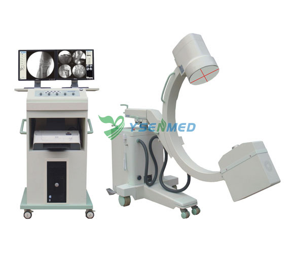
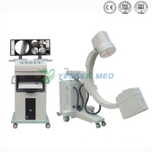
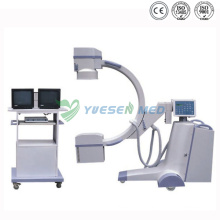
Medical High Frequency X-ray Mobile Digital C-Arm Machine


Medical High Frequency X-ray Mobile Digital C-Arm Machine
| Min. Order: | 1 |
|---|
| Place of Origin: | China |
|---|---|
| Supply Ability: | 500 Sets/Year |
| Certificate: | ISO13485 |
Basic Info
Model No.: YSX-C35D
Product Description
Model NO.: YSX-C35D Type: X Ray Equipment Group: All Double Focus Fixed Anode Focus:1.5mm / 0: Anode Focus:1.5mm / 0 Image Intensifier: Toshiba 9"(Including 3 Fields of View 4.5"/ 6"/9") Mas: 1mA.S-250mA.S Digital Radiography (Digital Spot Film): 40kv-120kv;Max 16mA Display: Display2 17"TFT-LCD Display Specification: ISO certificate HS Code: 9022199090 Classification: Imaging Diagnostic Equipment Certification: ISO13485 Power: 3.5kw High Voltage X-Ray Generator: Working Frequency:40kHz Tube Voltage: Fluoroscopy Radiograph:40kv-110kv General Radiography: 40kv-120kv;1.0mA.S-250mA.S Image Resolution Index: Grey Level:10 Resolution:22 Lp/Cm Trademark: YSENMED Origin: China High frequency Mobile Digital C-Arm X-Ray Machine (YSX-C35D)
Main componentsTechnique parameter
IMD X-ray tubeModel: YSX0705Model: YSX0706
Power: 3.5kWPower: 5.0kW
Double focus fixed anode focus: 1.5mm / 0.6mmDouble focus rotational anode focus: 0.3mm / 0.6mm
Anode Heat Capacity: 40kHu X-ray tube sleeve heat capacity: 667kHuAnode Heat Capacity: 200kHu X-ray tube sleeve heat capacity: 800kHu
High Voltage X-ray GeneratorWorking frequency: 40kHz
Image intensifier9"(including 3 fields of view 4.5"/ 6"/9")
Tube voltageFluoroscopy Radiograph: 40kV-110kVFluoroscopy Radiograph: 40kV-120kV
Tube currentFluoroscopy: 0.5-4mA Enhanced Automatic Fluoroscopy: 1mA-8mA Pulse Fluoroscopy: 2pps; 8mA
MAs1mA. S-250mA. S
Fluoroscopy modeManual Fluoroscopy; Semi-Automatic Fluoroscopy; Automatic Fluoroscopy; Enhanced Automatic Fluoroscopy; Pulse Fluoroscopy
Intelligent exposure controlNo matter the object is in the center or on the fringing field of the image intensifier, the images are the same. The exposure dose is reduced.
Iris beam limiting devicewhen not exposure, keep in the state of Iris Beam limiting preview, when exposure, keep blanking cycle track
General radiography40kV-120kV; 1.0mA. S-250mA. Sstep length 1 kV; 1.0mA. S
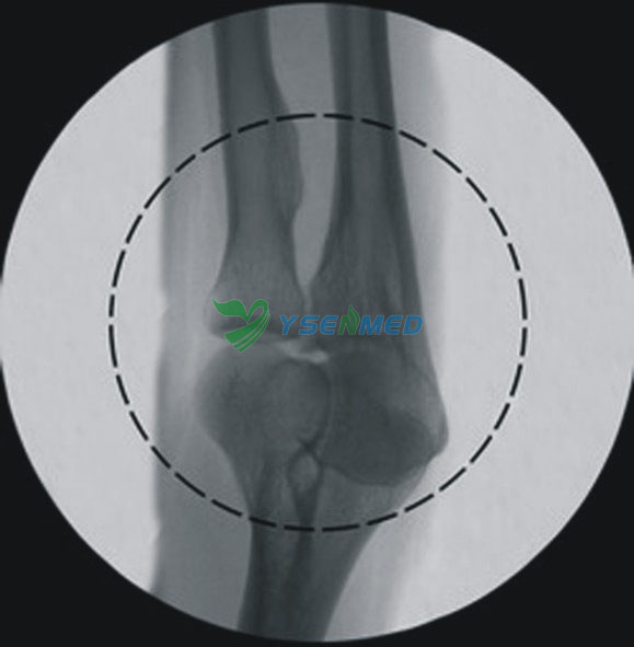
Digital radiography (digital spot film)40kV-120kV; Max 16mACommon screen film radiography is eliminated. Digital radiography can get better images
C-arm machinery indexC-arm stander(with perfect C arm balance system) of ultra-quiet motor-driven up and down, easy and handy to operate. The machine can maintain balance with up and down of the C arm.
Distance between focus and window>900mm, arc depth>650mm, horizontal moving range: 200mm vertical moving range: 400mm
Tilt angle: ± 12.5° Rotation: ± 180° Corner: 125° (-35° -- +90° )
Laser OrientationLaser positioning function can help to position precisely and reduce exposure times in the operation, reduce unnecessary radiation.
Radiography mode
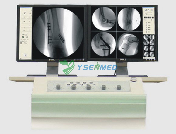
Image intensifier & Real-time digital image(1.3 million pixel) processing system
(CCD)Digital video cameramillion pixels high-definition, high-speed CCD camera, a 1024 × 1024 matrix, 30 frames per second image acquisition, through real-time adjustment of window width, window level, noise reduction, sharpening and Gain adjustment to ensure high-quality and high-resolution images.
Image resolution indexGrey level: 10 Resolution: 22 lp/cm
Camera virtual rotation designNo X-ray needed, also called digital image rotation, display the right angle when exposure, superior to camera mechanical rotation
Image processing system1. Real time edge enhancement(sharpening)Image sharpening is also called edge enhancement, that is, to make blurry edge of the image clearer
2. Real time adjustment of the window width and window levelObtain images by real-time adjustment of window width, window level, displaying images with different brightness.
3. Real time(static)multiform noise reductionNoise reduction, and improve image clarity
4. Real tim GAIN adjustmentCan amplify image signal, reduce x-ray dose, suitable for patient with super large size.
5. Real tim dynamic brightness compensation and R calibration functionsThrough dynamic logarithmic transformation, linear reduction the system signals and r calibration technology, dark parts of the image can be enhanced and clear rich-layer images can be obtained
6. Image conversing, up-down and right-left rotating, and frame freezing
7. Patient information management, Clinic report and print
8. The Image can be storage as JPG and DICOM 3.0 format, can be linked to hard-disk, USB and DVD and printer, meet the need of sharing the network information, communication and storage in hospital
DisplayDisplay2 17' TFT-LCD display
PrinterPrinter Laser / jet Printer
Contact us if you need more details on X-ray Machine. We are ready to answer your questions on packaging, logistics, certification or any other aspects about X-Ray Equipment、Medical Equipment. If these products fail to match your need, please contact us and we would like to provide relevant information.

Main componentsTechnique parameter
IMD X-ray tubeModel: YSX0705Model: YSX0706
Power: 3.5kWPower: 5.0kW
Double focus fixed anode focus: 1.5mm / 0.6mmDouble focus rotational anode focus: 0.3mm / 0.6mm
Anode Heat Capacity: 40kHu X-ray tube sleeve heat capacity: 667kHuAnode Heat Capacity: 200kHu X-ray tube sleeve heat capacity: 800kHu
High Voltage X-ray GeneratorWorking frequency: 40kHz
Image intensifier9"(including 3 fields of view 4.5"/ 6"/9")
Tube voltageFluoroscopy Radiograph: 40kV-110kVFluoroscopy Radiograph: 40kV-120kV
Tube currentFluoroscopy: 0.5-4mA Enhanced Automatic Fluoroscopy: 1mA-8mA Pulse Fluoroscopy: 2pps; 8mA
MAs1mA. S-250mA. S
Fluoroscopy modeManual Fluoroscopy; Semi-Automatic Fluoroscopy; Automatic Fluoroscopy; Enhanced Automatic Fluoroscopy; Pulse Fluoroscopy
Intelligent exposure controlNo matter the object is in the center or on the fringing field of the image intensifier, the images are the same. The exposure dose is reduced.
Iris beam limiting devicewhen not exposure, keep in the state of Iris Beam limiting preview, when exposure, keep blanking cycle track
General radiography40kV-120kV; 1.0mA. S-250mA. Sstep length 1 kV; 1.0mA. S
Digital radiography (digital spot film)40kV-120kV; Max 16mACommon screen film radiography is eliminated. Digital radiography can get better images
C-arm machinery indexC-arm stander(with perfect C arm balance system) of ultra-quiet motor-driven up and down, easy and handy to operate. The machine can maintain balance with up and down of the C arm.
Distance between focus and window>900mm, arc depth>650mm, horizontal moving range: 200mm vertical moving range: 400mm
Tilt angle: ± 12.5° Rotation: ± 180° Corner: 125° (-35° -- +90° )
Laser OrientationLaser positioning function can help to position precisely and reduce exposure times in the operation, reduce unnecessary radiation.
Radiography mode
Image intensifier & Real-time digital image(1.3 million pixel) processing system
(CCD)Digital video cameramillion pixels high-definition, high-speed CCD camera, a 1024 × 1024 matrix, 30 frames per second image acquisition, through real-time adjustment of window width, window level, noise reduction, sharpening and Gain adjustment to ensure high-quality and high-resolution images.
Image resolution indexGrey level: 10 Resolution: 22 lp/cm
Camera virtual rotation designNo X-ray needed, also called digital image rotation, display the right angle when exposure, superior to camera mechanical rotation
Image processing system1. Real time edge enhancement(sharpening)Image sharpening is also called edge enhancement, that is, to make blurry edge of the image clearer
2. Real time adjustment of the window width and window levelObtain images by real-time adjustment of window width, window level, displaying images with different brightness.
3. Real time(static)multiform noise reductionNoise reduction, and improve image clarity
4. Real tim GAIN adjustmentCan amplify image signal, reduce x-ray dose, suitable for patient with super large size.
5. Real tim dynamic brightness compensation and R calibration functionsThrough dynamic logarithmic transformation, linear reduction the system signals and r calibration technology, dark parts of the image can be enhanced and clear rich-layer images can be obtained
6. Image conversing, up-down and right-left rotating, and frame freezing
7. Patient information management, Clinic report and print
8. The Image can be storage as JPG and DICOM 3.0 format, can be linked to hard-disk, USB and DVD and printer, meet the need of sharing the network information, communication and storage in hospital
DisplayDisplay2 17' TFT-LCD display
PrinterPrinter Laser / jet Printer


| Main components | Technique parameter | ||||
| IMD X-ray tube | Model:YSX-C35D | Model:YSX-C50D | |||
| Power:3.5kW | Power:5.0kW | ||||
| Double focus fixed anode focus:1.5mm / 0.6mm | Double focus rotational anode focus: 0.3mm / 0.6mm | ||||
| Anode Heat Capacity:40kHu X-ray tube sleeve heat capacity:667kHu | Anode Heat Capacity:200kHu X-ray tube sleeve heat capacity:800kHu | ||||
| High Voltage X-ray Generator | Working frequency:40kHz | ||||
| Image intensifier | 9"(including 3 fields of view 4.5"/ 6"/9") | ||||
| Tube voltage | Fluoroscopy Radiograph:40kV-110kV | Fluoroscopy Radiograph:40kV-120kV | |||
| Tube current | Fluoroscopy: 0.5-4mA Enhanced Automatic Fluoroscopy: 1mA-8mA Pulse Fluoroscopy :2pps;8mA | ||||
| mAs | 1mA.s-250mA.S | ||||
| Fluoroscopy mode | Manual Fluoroscopy; Semi-Automatic Fluoroscopy;Automatic Fluoroscopy; Enhanced Automatic Fluoroscopy; Pulse Fluoroscopy | ||||
| Intelligent exposure control | No matter the object is in the center or on the fringing field of the image intensifier, the images are the same. The exposure dose is reduced. | ||||
| Iris beam limiting device | when not exposure, keep in the state of Iris Beam limiting preview ,when exposure,keep blanking cycle track | ||||
| General radiography | 40kV-120kV;1.0mA.s-250mA.s | step length 1 kV;1.0mA.s | |||
| Digital radiography (digital spot film) | 40kV-120kV;Max 16mA | Common screen film radiography is eliminated. Digital radiography can get better images | |||
| C-arm machinery index | C-arm stander(with perfect C arm balance system) of ultra-quiet motor-driven up and down, easy and handy to operate. The machine can maintain balance with up and down of the C arm. | ||||
| Distance between focus and window>900mm, arc depth>650mm, horizontal moving range :200mm vertical moving range:400mm | |||||
| Tilt angle :±12.5° rotation :±180°corner:125°(-35°-- +90°) | |||||
| Laser Orientation | Laser positioning function can help to position precisely and reduce exposure times in the operation, reduce unnecessary radiation. | ||||
| Radiography mode | |||||
| Image intensifier & Real-time digital image(1.3 million pixel) processing system | |||||
| (CCD)Digital video camera | million pixels high-definition, high-speed CCD camera, a 1024 × 1024 matrix, 30 frames per second image acquisition, through real-time adjustment of window width, window level, noise reduction, sharpening and Gain adjustment to ensure high-quality and high-resolution images. | ||||
| Image resolution index | Grey level:10 Resolution:22 lp/cm | ||||
| Camera virtual rotation design | No X-ray needed, also called digital image rotation,display the right angle when exposure, superior to camera mechanical rotation | ||||
| Image processing system | 1.Real time edge enhancement(sharpening) | Image sharpening is also called edge enhancement, that is, to make blurry edge of the image clearer | |||
| 2.Real time adjustment of the window width and window level | Obtain images by real-time adjustment of window width,window level, displaying images with different brightness. | ||||
| 3.Real time(static)multiform noise reduction | Noise reduction, and improve image clarity | ||||
| 4.Real tim GAIN adjustment | Can amplify image signal, reduce x-ray dose, suitable for patient with super large size. | ||||
| 5.Real tim dynamic brightness compensation and R calibration functions | Through dynamic logarithmic transformation,linear reduction the system signals and r calibration technology, dark parts of the image can be enhanced and clear rich-layer images can be obtained | ||||
| 6.Image conversing ,up-down and right-left rotating,and frame freezing | |||||
| 7.Patient information management ,Clinic report and print | |||||
| 8.The Image can be storage as JPG and DICOM 3.0 format, can be linked to hard-disk ,USB and DVD and printer,meet the need of sharing the network information ,communication and storage in hospital | |||||
| Display | Display2 17' TFT-LCD display | ||||
| Printer | Printer Laser / jet Printer | ||||
| Standard configuration | 1.Combined tube(5.0kW)IMD | 1 set; | |||
| 2.Image intensifier (9 inch 3 fields of view) | 1 set | ||||
| 3.C-arm stander (with full C-arm balance system) | 1 set; | ||||
| 4.Exported grid | 1 set | ||||
| 5.Laser positioning | 1 set; | ||||
| 6.1.3million pixel 1024×1280×10bit camera | 1 set; | ||||
| 7.Electric adjustable beam limiting device | 1 set | ||||
| 8.17 inch LCD high resolution screen | 2 sets | ||||
| 9.Real-time digital imageprocession system (station) | 1 set; | ||||
| 10.Printer | 1 set | ||||
Contact us if you need more details on X-ray Machine. We are ready to answer your questions on packaging, logistics, certification or any other aspects about X-Ray Equipment、Medical Equipment. If these products fail to match your need, please contact us and we would like to provide relevant information.
Product Categories : Medical X-ray Machine > C arm
Premium Related Products

Other Products
Hot Products
Laboratory equipments portable blood gas analyzer/blood gas and electrolyte analyzer MSLAB18iMSLPO-A Finger Pulse Oximeter handheld pulse oximeter in GuangzhouFDA CE Certificate MT01A Hospital Medical Crash Trolley / CartFactory price professional portable echo ultrasound priceMSLER02 Elisa Microplate Washer and Microplate Washer for Microplate Reader VersamaxElisa Microplate Reader MSLER01-Microplate Reader Function In MSLER01Human & Animal use Portable ultrasound machine MSLPU03-MMSLCU18-2015 New and best 3D&4D portable ultrasound machine used in Human and vetLatest Medical Full-auto blood Hematology analyzer with CE ISO Certificates plus Manufacture priceUrine analysis equipment | Urine test machine - MSLUA02Cheap and new dental chair /dental unit price (MSLDU01-M)Exquisite packaging: Handheld veterinary ultrasound scanner used in bovine, sheep, etc (MSLVU04M))Semi-auto Blood Coagulation Analyzer with CE Approved(MSLBA18)MSLLR01 - Medical Lead Rubber SheetAutomated chemistry analyzers(MSLAB07)(MSLAB01)Cheap Portable sysmex Clinic Auto veterinary Hematology blood analyzer